Synonyms.—Rheumatoid Arthritis; Rheumatic Gout.
Definition.—A chronic inflammatory disease of the articulations, characterized by progressive changes in the joint structures, and with periarticular formation of bone, causing marked deformity and greatly impairing their function.
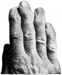
Etiology.—There appears to be a wealth of theory, and a dearth of facts, as to the etiology of this disease. For years it was regarded by many as a phase of rheumatism, or a near relative, while others were equally positive that its similarity to gout 'was a sufficient proof that they were of one and the same family. More recent observers have declared their belief that the disease is entirely distinct from either; in fact, Heberden, in 1804, declared his belief in a distinct lesion, and insisted that there should be a divorce from gout and rheumatism. He first described the "small nodular outgrowths upon the terminal joints of the fingers," and which are now universally referred to as Heberden's nodes.
Fuller and Garrod, early converts to this belief, called attention to the fact that in arthritis deformans there were not the blood changes nor visceral manifestations that there were in rheumatism, nor could there be detected a trace of urates in this disease, while in gout it was ever present.
The neurotrophic origin of the disease has many followers, the profession's attention having been directed to this theory by the writings of the Mitchells, father and son, in America; Weichman, in Germany; and Ord and Duckworth, in England.
Hutchinson believes that it is neither a distinct disease from rheumatism, nor a variety, but a blending of the two, or a hybrid. The latest theory is that of infection, and that the cause is microbic. The etiology of the disease is thus seen to be doubtful, to say the least.
Predisposing Causes.—Dr. Garrod's contribution to the "Twentieth-Century Practice" contains a very interesting and instructive article on this disease, and from a tabulated report of five hundred cases the following factors figure as predisposing causes:
Age.—The greatest number of cases occur between the ages of forty and fifty-five, though no age enjoys immunity, three cases occurring before the age of ten, and one between the ages of eighty and ninety; there were less than fifty cases occurring under twenty-five years of age.
Sex.—Of the five hundred cases, four hundred and eleven were women, leaving only eighty-nine cases among the men.
Hereditary Predisposition.—In two hundred and sixteen cases the family history revealed lesions of the articulations. One woman's history revealed the fact that her father and mother had joint affections, and that of six living children, her three brothers and two sisters suffered from enlargement of the joints; thus the entire family of eight were victims of articular deformity.
Rheumatism and Gout.—In one-third of all the cases reported, gout held a prominent place, while rheumatism occurred in but sixty-four cases.
Exposure, dietetic errors, worry, care, and injuries have all been regarded as predisposing causes to the disease, though a reference to his tabulated report would not suggest them as playing an important part.
Pathology.—In the pathological changes that take place we notice, first, that there are no deposits of urate of soda as in gout, and the extensive structural changes that take place in the joints are not found in rheumatism. There may be effusion in the early stages, but as progressive changes take place, this disappears, and the first effects seem to be felt in the cartilage.
Proliferation of cells takes place, fibrillation follows, which results in softening of the cartilage, especially in the center where the circulation is feeble, and the friction great, from the opposing ends of the bones; this results in the center of the cartilage disappearing, and the exposed ends of the bones, from friction, become polished like ivory, and are termed eburnated. The outer portion of cartilage does not share in the destruction which takes place in the center, owing to absence of pressure, and instead of thinning we find enlargement; finally, ossification takes place, forming the so-called osteophytes, which often cause a locking of the joints.
In addition, nodes may develop from the periosteum along the shaft of the bone. Following these changes, inflammation of the synovial membrane takes place, exudation results, which may become organized, and, in rare cases, ossification takes place. Finally the capsule and ligaments become thickened, resulting in ankylosis. After this, atrophy of the muscles may follow, and, still more rarelv, neuritis. The joint turns outward to the ulnar side: the same changes sometimes take place in the toes, they likewise turning outward.
 Symptoms.—The dividing line between the acute, subacute, and chronic, or "Heberden's nodes," and the "general progressive form," requires greater skill than most practitioners possess; for the division is more technical than real, all cases being more or less chronic.
Heberden's Nodes.—In this form the chief characteristics are the nodosities that are formed, osteophytes, at the sides of the distal phalanges, and occur far more frequently in women than in men, usually between the age of thirty and forty. It is more apt to follow rapid child-bearing or undue lactation. The menopause is also a fruitful time for their development. They are apt to be preceded by rheumatism or gout, though not necessarily, for the disease is entirely distinct.
The patient notices that the joints are swollen, slightly reddened and tender on motion, or, if struck, sometimes they are quite painful, though this is exceptional. There may come a period of intermission, and the disease appears to have subsided, but only to reappear, the enlargement gradually increasing, till the knotty excrescences prevent the motion of the joints, and they become locked.
In time the cartilage gives way, and crepitus can be distinguished, which is followed by eburnation. The general health is but little affected in many cases, though some are attended by gastric disturbances and anemia.
General Progressive Form.—This may occur in the acute and chronic form; when acute, it may simulate acute articular rheumatism, the patient having slight fever, with swelling of the joints, the synovial sheaths, and bursse. There is little redness, however, and usually not a great deal of pain. After a time the disease is stayed, sometimes for years, when some aggravation, such as child-bearing, starts anew the fires that were supposed to be extinct, and the joints take^on the usual characteristics.
The Chronic Form is the one usually found. It comes on slowly and insidiously, first in one or two joints, then in the corresponding ones on the opposite member, until the entire articulations are involved; the articulation of the hands being affected more frequently than any other joint, though none are exempt.
The first symptom is a slight swelling of the joint, attended with some stiffness and pain. There may be effusion into the joint, with swelling of the sheaths and bursse. There is often but little pain, sometimes none, but in very rare cases the pain is excruciating. As in the other forms, there are times when the disease seems to be stayed, and then is renewed, each time resulting in greater joint changes, till finally the articulation, from osteophytes, thickening of sheaths, and atrophy of muscles, is greatly deformed.
In the hands, the joints are turned outwards or to the ulnar side, and the same may be said when the toes are affected, sometimes the phalanges overlapping. The disease may be confined to the hands, or be followed by the knees, ankles, hips, and vertabrae, continuing till every articulation becomes involved, and the patient, drawn and weak with suffering, becomes perfectly helpless.
Diagnosis.—In the advanced stage there is but little difficulty in making a diagnosis; the great deformity, with but little pain, the turning outward of the fingers and toes, and the immobile condition of the joints, certify to the trouble. In the earlier stages, however, it may be mistaken for chronic rheumatism; but even here the history, with the more gradual invasion, will help to distinguish the one from the other.
Prognosis.—This is unfavorable if the disease is well established, especially where the joints are locked from bony deposits. In the earlier stages it may be arrested, if not completely cured. The disease is rarely ever fatal.
Treatment.—The physician usually sees these cases in the advanced stage, after two or more joints are affected and after ankylosis is partially established; hence the treatment is not satisfactory, but little, if any, benefit resulting from medication; gradually the deformity increases until the patient is a helpless cripple.
If seen early, we may hope to benefit our patients. It is very important to correct any uterine or rectal troubles that may exist, before administering remedies internally. All sources of nerve impingement, whereby capillary circulation is impaired, must be removed, and any systemic wrongs corrected. Berberis aquifolium, stillingia, bryonia, potassium iodide, and the salicylates will be followed by improvement in the earlier stages. Massage is of greater importance, and should be used faithfully and continuously. Where the patient has means, a course at Hot Springs, Virginia, or Arkansas, will be of benefit.
The diet should be generous, but of such foods as will give increased nourishment. Exercise in the open air should be taken; in fact, hygienic measures will form a very important part of the treatment. Where the patient is unable to visit the famous Springs, some benefit will result from the hot vapor-bath. As a last resort the surgeon may have to be called to our assistance to remove deformities.
The Eclectic Practice of Medicine, 1907, was written by Rolla L. Thomas, M. S., M. D.

