Synonym.—Biliary Calculi; Cholelithiasis.
Definition.—Concretions, which form in the biliary passages or gall-bladder.
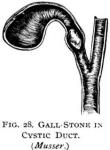
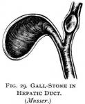
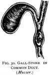
Etiology.—It is not definitely known as to the positive cause or causes that give rise to gall-stones. They are found within the hepatic duct and the gall-bladder, and rarely in the cystic duct. They occur more frequently after the age of sixty, although they have been found in infancy and early life. According to Naunyn, they occur far more frequently in women than in men, about four to one, especially in women who have borne children. Tight lacing, sedentary habits, constipation, and fatty and starchy food also predispose to gall-stones.
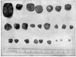 The chief constituents of gall-stones are cholesterin and pigment-lime precipitated from the bile, as the result of the decomposition of such salts as hold them in solution. The more recent theory is that they are the result of micro-organisms. The fact that the gall-bladder is the habitat for the colon bacilli, streptococci, staphylococci, pneumococci, and the typhoid bacilli, and that gall-stones are frequently associated with the infectious fevers, and the experiment of Gilbert and Fournier, who produced gall-stones in animals by injecting micro-organisms into the gall-bladder, give weight to the theory.
Composition and Appearance.—Gall-stones vary in size and number, shape, color, formation, and consistency. They are composed of cholesterin bile-pigment, especially bilirubin, the lime-salts, and rarely phosphorus, magnesia, with occasional traces of iron and copper. Their consistency depends upon their constituents; thus, when made up of cholesterin and mucus, they are soft, and may be cut like wax, while those in which the lime-salts are well represented are hard and brittle. The color varies from white, the cholesterin stones, to the yellow, dark-brown, or almost black, depending upon the amount of bile-pigment present.
There may be but one stone, or there may be thousands. Otto recording a case where there were seven thousand eight hundred and two stones in a single case. The fewer the stones the larger they become, and where only one exists it may attain a size of four or five inches in length. The single stone is usually found in the gall-bladder, while the smaller ones may be found anywhere in the biliary tract, even to the minutest bile-duct. The minute stones are sometimes called gall-sand, and no doubt the great number recorded by Otto were of this kind.
Their shape depends upon their consistency as well as number. When soft, they may be flattened, and when hard, they may contain facets when crowded together; others are oblong, oval, or egg-shaped, depending upon pressure against each other. On section, the stone often shows a nucleus of cholesterin, the remainder being made up of concentric layers, the outer layers containing the various salts, being brown and hard.
The ducts, where the stone is located, may be dilated or saccular, and the mucous membrane smooth; or, if inflammation has been set up, it is thickened or ulcerated. In the latter case, adhesions to other parts take place. Perforation may occur into the peritoneal cavity or into other organs, and fistulous tracts may be established into the bowel, kidney, ureter, stomach, bronchi, or abdominal wall, the stones being discharged in this way. Suppurative inflammation may result in any of the ducts where the calculi may be located, empyemia resulting.
Symptoms.—Gall-stones may be present for a long time in the gall-bladder, and finally pass out into the bowel without any symptoms, the presence of the stones in the stool being the first knowledge of there having been biliary calculi. A case of this kind came under my notice where the patient passed a handful of stones the size of filberts. Occasionally there will be an uneasy sensation in the right hypochondriac region, especially on change of position, when the stones remain in the gall-bladder; generally, however, the first evidence of hepatic calculi is when the stone starts on its journey for the bowel, and manifests itself in a sudden, intense, and lancinating pain in or near the region of the gall-bladder—hepatic colic.
The pain, beginning in the right hypochondrium or epigastrium, radiates in every direction, especially upwards in the right thorax, extending to the back and right shoulder. The attack occurs most frequently in the after part of the day or about midnight. It may last but a few minutes and cease, or for hours, days, and even weeks. As soon as the stone reaches the intestine the pain suddenly ceases. During the paroxysm of pain the patient may roll on the floor, flex the right limb, and grasp the abdominal wall for relief. The excruciating pain causes him to cry out with the suffering, the face becomes pallid, and frequently a cold sweat bathes the patient. Vomiting of bile is a common symptom.
The pulse is slow and feeble, or small and rapid. Rigors are often present, followed by fever, the temperature rising to 102° or 103°, and in rare cases going to 104° or 105°. The pain is intermittent in character, subsiding for a few minutes, only to appear with apparent redoubled force.
In from eight to twenty-four hours jaundice makes its appearance, although it does not occur in all cases. When the paroxysms have been unusually severe, the patient is completely exhausted at their termination, although the strength is rapidly renewed.
During an attack the liver is somewhat enlarged, and may be felt several inches below the costal line. There is tenderness in this region. In some cases there is enlargement of the spleen. Where the abdominal walls are thin, the presence of calculi can sometimes be determined by palpation.
Dropsy of the gall-bladder—hydrop vesicae felleae—may arise when the stone lodges in the cystic duct; giving rise to chronic obstruction.
Empyemia of the gall-bladder is a rare complication, the organ becoming greatly distended with pus.
In very rare cases, rupture of the duct occurs, followed by fatal peritonitis.
Diagnosis.—This is comparatively easy, the sudden paroxysms of pain in the epigastric region extending to the right shoulder, vomiting, great prostration, slow pulse, clammy skin, and jaundice, make a picture that can scarcely be mistaken for appendicitis, renal, or lead colic. The positive diagnosis is, of course, made when the stone is passed in the stools, which should carefully be washed through a sieve.
Prognosis.—The prognosis of the individual attack is usually favorable, although death has occurred in a first paroxysm, where the vitality was low, the result of fatty heart. Cerebral hemorrhage, the result of an attack, has-also proved fatal, while a local peritonitis, the result of perforation of the duct, has terminated fatally. Empyemia of the gall-bladder, attended by septic fever, carcinoma, and cardiac degeneration, render the prognosis very grave.
Treatment.—Our first object is to give relief to the excruciating pains of the distressed patient. This will be accomplished by inhalations of chloroform, and the hypodermic use of morphine. Most benefit will result from hot fomentations or the sitz-bath, or the old alcohol sweat. While the paroxysm is on, relief may be had by placing a napkin, moistened with chloroform, over the painful locality; but care must be used, and the cloth removed every few seconds, or the part will be burned.
Internally, large doses of dioscorea in hot water affords some relief; say dioscorea fifteen or twenty drops in a fourth of a cup of hot water every hour. The earlier Eclectics prescribed lobelia and asclepias in infusion, till diaphoresis and relaxation were fully established; although unpleasant, it is still good treatment.
As soon as the stone passes into the intestine, a full dose of antibilious physic and cream of tartar should be given, to remove, not only the calculi, but accumulated bile and fecal matter. Following an attack, remedies should carefully be selected to prevent further formations of the concretions, if possible.
Chionanthus, chelidonium, leptandrin, hydrastin, and podophyllin, as indicated, should be given for several weeks. The sodium salts should also be given freely. Olive-oil, an ounce, night and morning, is an old remedy, and may do some good. Sodium phosphate and Podophyllin are not to be forgotten, as they are among our most reliable agents in this affection.
A very important part of the after treatment is the hygienic and dietetic. The patient should take regular exercise daily in the open air; horseback-riding is preferable, where the patient is able to profit by such advice.
The diet should be largely vegetarian, although lean meats and fish may be allowed. All starchy and fatty foods, as well as sugar and pastries, should be avoided. Green vegetables, fruits, and skimmed milk, whey, 'or buttermilk should be the chief diet.
The bowels should never be allowed to become constipated, and if the soda salts are taken night and morning, there will be no danger from this source. Where the stone becomes permanently lodged and no relief from pain or soreness follows appropriate treatment, and when a septic fever with jaundice arises, which tells of pus in the gall-bladder, the case should be placed in the hands of the surgeon for operative treatment.
The Eclectic Practice of Medicine, 1907, was written by Rolla L. Thomas, M. S., M. D.
